Is My Lump Cancerous?

The Confirmatory Tests

Is my breast lump
cancerous?

A question which gives sleepless nights to the family. And unmeasureable levels of anxiety.
So, after the examination, the next step now is to confirm, if this is really a cancer. And why is it necessary to confirm? Because, it could very well be non cancerous. There are some conditions, in which, the lump in the breast feels like a cancer, but is not - Granulomatous Mastitis, Infections etc. Or it could very well be one of those non cancerous tumours in the breast like fibroids etc. In most cases, I will definitely have a rough idea during examination itself, as to whether it is cancerous or not. For the confirmation part, we need to do two tests:

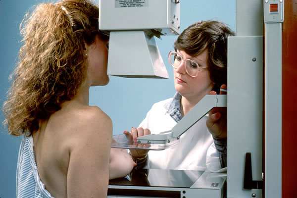
Mammography : To locate the tumour
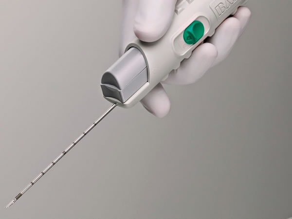
Core Biopsy : To confirm the cancer

Mammography

Mammography is a special X Ray of the breast in two directions. In addition to X Ray, an ultrasound of the breast is also done, and the Radiologist co relates the finding of the X Ray and the ultrasound and gives the report. (With newer techniques now, instead of a simple X Ray, a Tomography is done. In selected patients, we also do an MRI).

Mammography tell us about the exact location, size and nature of the tumour.

It also tells us the following:

Is it a 'single' tumour? or there are more than one? (This point will influence the choice of surgery)
What is the chance that this tumour (or tumours) is a cancer?

To answer the second question, the final report of mammography will contain a grading called "BIRADS" to tell us what does the Radiologist feel after doing Mammography. There are 6 categories:

BIRADS 1 and 2 are non cancerous.
BIRADS 3 - it is not clear, and a repeat mammography will be advised in three to six months.
BIRADS 4 - there is suspicion of cancer.
BIRADS 5 - we are almost sure of cancer.
BIRADS 6 - is written when there is a proven cancer.

Mammography for breast lumps
Core Biopsy Needle for Breast tumour

Core Biopsy

Core biopsy is the confirmatory test for breast cancer. It may be done by a Radiologist, where is it usually an ultrasound guided biopsy or a vacuum biopsy for tumours which are not felt by hand. Or in cases where the 'lump' can be 'felt' clearly by hand, it may be done by the Surgical Oncologist. A Core Biopsy not only confirms the cancer, but also tells us which 'type' it is

Core biopsy is an OPD based procedure. It is done under 'local anesthesia', where a small injection is given over the area of lump to make it numb, and then, a special needle (shown in the picture) is inserted into the lump and few pieces of the lump pulled out. The pieces are preserved in a solution and sent to a pathologist for testing.

So what will the reports show?

A Mammography report of a typical breast cancer patient will describe the tumour and in the end, menton the BIRADS category - in most cases, BIRADS 5, and in some, it may mention BIRADS 4A or 4B or 4C. And the biopsy report gives us the exact type of tumour. The following are a sample of words which can be found in the biopsy report of a typical cancer patient:

Infiltrating (Invasive) Duct Carcinoma
Infiltrating (Invasive) Lobular Carcinoma
Ductal Carcinoma in Situ, No invasive element seen
Ductal Carcinoma in Situe, micro invasion seen
In addition to one of the above, there will be a separate mention of 'Immuno Histo Chemistry (IHC)' which will tell us about the type of breast cancer this is. There are quite a few markers of IHC which we do for each and every breast cancer patient, the three most important being - ER (Estrogen Receptor), PR (Progesterone Receptor) and HER2neu (or CerbB2)

Please remember, these are just examples to help you understand. There are lots of more terms to be described, but we don't want to go too much into it. Please ask your Oncologist to help you decipher terms in the report.

Our Approach at this Step


After the Core Biopsy report comes, I assess it in detail, sit with the patient and the relatives, and explain to them line by line. I tell them that the cancer is indeed confirmed (or as the case may be), I place my hand on hers and assure her - 'Breast cancer treatment is far more advanced as compared to before. We are together in this and I shall guide you and walk you through each step of this journey'. We now go on to the Staging of cancer.


Click the buttons below for the Next topic or the Previous topic, or go to topic of your choice by using the menu below


TOPICS IN THIS SECTION

All the links are in blue colour. You can click the links to go to that page. The page you are presently reading is pink in color


Journey of BC: Diagnosis


1. I feel a lump! - The first visit to doctor
2. Is it really cancer? - Confirmatory tests - You are presently on this page
3. What is the stage? - Staging and fitness tests
4. Will it be surgery first? or chemo first? - Treatment planning and sequence

Journey of BC: Surgery


5. Should I conserve or remove breast? - Choice of Surgeries for breast cancer
6. How long will be the surgery? - Admission and Surgery
7. Care after surgery - Precautions and guidelines
8. What next? - The Pathology Report

Journey of BC: Further Treatment


9. How many chemo cycles? - Chemotherapy Consultation
10. Is a PORT necessary? - ChemoPORT insertion
11. Will I be normal during chemo? - The Chemotherapy time
12. Is Radiation painful? - Radiation Therapy
13. Yes!! I did it! - Treatment is over
14. How frequently do I meet doc? - Follow up guidelines

Other Topics


Risk Factors - The Risk Factors for Breast Cancer
Symptoms of Breast Cancer - Know the Symptoms of Breast Cancer
Early Detection of Breast Cancer - The Guidelines

Neo Adjuvant Chemotherapy (NACT) - For LABC
Sentinel Node Biopsy - How is it done?
Targeted Therapy - Trastuzumab
Hormone Therapy - Who gets it?